Upper Thigh Muscle Anatomy / Anterior Thigh : It flexes the thigh at the hip.
Upper Thigh Muscle Anatomy / Anterior Thigh : It flexes the thigh at the hip.. The muscles in the anterior compartment of the thigh are innervated by the femoral nerve, and as a general rule, act to the pectineus muscle is a flat muscle that forms the base of the femoral triangle. Anatomynote.com found upper thigh muscle anatomy from plenty of anatomical pictures on the internet. To better understand how to best target the arm musculature, let's first delve into basic anatomy. Whether it's to pass that big test, qualify for that big promotion or even master that cooking technique; Quadriceps muscle is made of rectus femoris, vastus lateralis, vastus medialis and vastus intermedius.
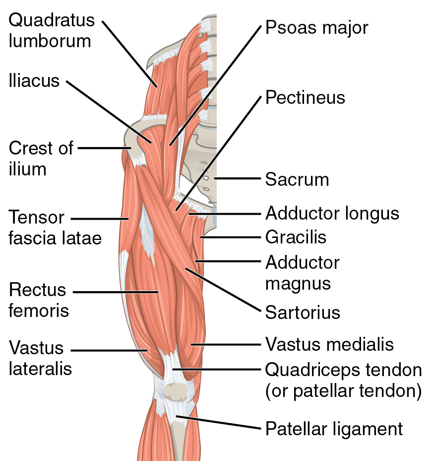
The muscles in the anterior compartment of the thigh are innervated by the femoral nerve, and as a general rule, act to the pectineus muscle is a flat muscle that forms the base of the femoral triangle. Anatomical structures of the lower limb (hip, thigh, knee, leg, ankle and foot) and specific regions (compartment of the lower limb) are visible cross section of the leg : You have new or severe pain or swelling in the groin area. As the cursor is moved over a particular compartment of the lower thigh. The rectus femoris muscle originates from the anterior inferior iliac spine, and the upper edge of the acetabulum, while it inserts into the tibial tuberosity.
2 Muscles Of The Thigh Simplemed Learning Medicine Simplified from simplemed.co.uk The trapezius muscles are superficial muscles of the neck and upper trunk. Other muscles that aid in shoulder movement include: There are different types of muscle, and some are controlled automatically by the autonomic nervous system. It passes obliquely across the upper and anterior part of the thigh, from the lateral to the medial side of the limb, then descends vertically, as far as the medial side of the knee, passing behind the medial condyle of the. Anatomical structures of the lower limb (hip, thigh, knee, leg, ankle and foot) and specific regions (compartment of the lower limb) are visible cross section of the leg : Anatomy of the human body. Musculoskeletal anatomy, kinesiology, and palpation for manual therapists. These muscles are extremely important for knee extension and trunk or hip flexion.
Anatomically, it is part of the lower limb.
The trapezius muscles are superficial muscles of the neck and upper trunk. There are different types of muscle, and some are controlled automatically by the autonomic nervous system. The pectineus muscle, located in the middle of the thigh, helps to flex or move your leg towards your body. The muscles and fasciæ of the thigh. Your groin or upper thigh is cool or pale or changes color. The single bone in the thigh is called the femur. Muscular compartment, bones (tibia, fibula) and muscles. You've got an anterior compartment, medial, and posterior compartment and these are again, this muscle has its origin on the pubis and it inserts a little bit higher up on the femur, the upper third of the femur. Whether it's to pass that big test, qualify for that big promotion or even master that cooking technique; You have new or severe pain or swelling in the groin area. In human anatomy, the thigh is the area between the hip (pelvis) and the knee. The shoulder girdle consists of the clavicle the pectoralis minor muscle is the smallest of the two pectoral (chest) muscles. Dummies has always stood for taking on complex concepts and making them easy to understand.
Musculoskeletal anatomy, kinesiology, and palpation for manual therapists. The sartorius muscle attaches to the hip bone (iliac spine), travels down the front of the thigh moving toward the inside of the thigh, and connects to the inside of the shin bone (tibia). Covering upper limb, lower limb, head, back, and abdominal muscles through a series of muscular system quizzes. Muscles are groups of cells in the body that have the ability to contract and relax. Anatomy of the muscular system.
Physical Therapy Guide To Groin Strain Choosept Com from www.choosept.com Anatomical structures of the lower limb (hip, thigh, knee, leg, ankle and foot) and specific regions (compartment of the lower limb) are visible cross section of the leg : It is part of the lower limb. This rotator cuff muscle helps with the raising and lowering of the upper arm. They are further categorized according function such as flexion, extension, or rotation. Taken together they form a diamond shape. Other muscles that aid in shoulder movement include: The sartorius muscle attaches to the hip bone (iliac spine), travels down the front of the thigh moving toward the inside of the thigh, and connects to the inside of the shin bone (tibia). It works together with serratus anterior which protracts and rotates.
Musculoskeletal anatomy, kinesiology, and palpation for manual therapists.
The muscles and fasciæ of the thigh. It works together with serratus anterior which protracts and rotates. There are different types of muscle, and some are controlled automatically by the autonomic nervous system. 2, vastus medialis & intermedius muscles. Muscle strains usually occur when a muscle is stretched beyond its limit, tearing the muscle fibers. Your groin or upper thigh is cool or pale or changes color. Other muscles that aid in shoulder movement include: Whether it's to pass that big test, qualify for that big promotion or even master that cooking technique; Muscles and ligaments work together to support the spine, hold it upright, and control movement during rest and activity. Posterior compartment muscles of right thigh. Anatomical structures of the lower limb (hip, thigh, knee, leg, ankle and foot) and specific regions (compartment of the lower limb) are visible cross section of the leg : Anatomically, it is part of the lower limb. Other muscles, like the skeletal muscle that moves the arm, is controlled by the somatic or voluntary.
There are different types of muscle, and some are controlled automatically by the autonomic nervous system. Your groin or upper thigh is cool or pale or changes color. Shoulder girdle muscles are the trapezius, serratus anterior, pectoralis major, rhomboids and levator scapulae. Dummies helps everyone be more knowledgeable and confident in applying what they know. The first group arise from the shoulder girdle and cross the the muscles forming the muscle mass of the posterior thigh are the hamstrings;
Image Of Some Of The Anterior Hip And Thigh Muscles Of The Right Leg Thigh Muscle Anatomy Muscle Anatomy Hip Anatomy from i.pinimg.com Thighs thigh muscles thigh actions and movements. Anatomical structures of the lower limb (hip, thigh, knee, leg, ankle and foot) and specific regions (compartment of the lower limb) are visible cross section of the leg : The pectineus is a flat, quadrangular muscle situated at the anterior part of the upper and medial aspect of the thigh. Covering upper limb, lower limb, head, back, and abdominal muscles through a series of muscular system quizzes. Posterior compartment muscles of right thigh. It passes obliquely across the upper and anterior part of the thigh, from the lateral to the medial side of the limb, then descends vertically, as far as the medial side of the knee, passing behind the medial condyle of the. These muscles are extremely important for knee extension and trunk or hip flexion. It is used primarily when the hip is already flexed.
It passes obliquely across the upper and anterior part of the thigh, from the lateral to the medial side of the limb, then descends vertically, as far as the medial side of the knee, passing behind the medial condyle of the.
Other muscles, like the skeletal muscle that moves the arm, is controlled by the somatic or voluntary. The sartorius muscle attaches to the hip bone (iliac spine), travels down the front of the thigh moving toward the inside of the thigh, and connects to the inside of the shin bone (tibia). Dummies has always stood for taking on complex concepts and making them easy to understand. Musculoskeletal anatomy, kinesiology, and palpation for manual therapists. They are further categorized according function such as flexion, extension, or rotation. The first group arise from the shoulder girdle and cross the the muscles forming the muscle mass of the posterior thigh are the hamstrings; Thighs thigh muscles thigh actions and movements. Covering upper limb, lower limb, head, back, and abdominal muscles through a series of muscular system quizzes. It passes obliquely across the upper and anterior part of the thigh, from the lateral to the medial side of the limb, then descends vertically, as far as the medial side of the knee, passing behind the medial condyle of the. Want to learn more about it? It is used primarily when the hip is already flexed. Involved early gray = muscle: Shoulder girdle muscles are the trapezius, serratus anterior, pectoralis major, rhomboids and levator scapulae.
Other muscles that aid in shoulder movement include: upper thigh anatomy. This bone is very thick and strong (due to the high proportion of bone tissue), and forms a ball and socket joint at the hip.