Hip And Leg Bone Diagram / Hip Anatomy : Bringing the leg back towards the midline.
Hip And Leg Bone Diagram / Hip Anatomy : Bringing the leg back towards the midline.. For example, the leg bones come together at the knee to form a hinge joint that enables the knee to bend back and forth. Download this free vector about diagram showing the hip bone treatment, and discover more than 11 million professional graphic resources on freepik. Нога (від стегна до ступні). Archeologists in belgium have discovered walls made. The hip bone (os coxae, innominate bone, pelvic bone or coxal bone) is a large irregular bone, constricted in the center and expanded above and below.
Body diagram was taken from the hip joint including the pelvis, upper body and the. Thigh (the part of the human leg between the hip and the knee). The knee joint is the largest joint in the body and is primarily a hinge joint, although some sliding and rotation occur. Tensor fascia lata trigger point in it band and hip pain dr perry details the tensor fascia late trigger point that cause hip pain and it band syndrome hip injuries hip disorders take a look at some mon and not so. Bones are mostly made of the protein collagen, which forms a soft framework.
Human Anatomy for the Artist: November 2011 from 1.bp.blogspot.com Clinically, hip dislocation presents itself with instability and limited abduction of the hip joint, and leg shortening with asymmetry of the gluteal folds. Hip and leg bone markings. The mineral calcium phosphate hardens this framework, giving it strength. This means that your feet. Standard radiography view of anatomical structures of the lower limb. The knee joint is the largest joint in the body and is primarily a hinge joint, although some sliding and rotation occur. The knee joint is the largest joint in the body and is primarily a hinge joint, although. The two bones beneath your knee that make up your shin are.
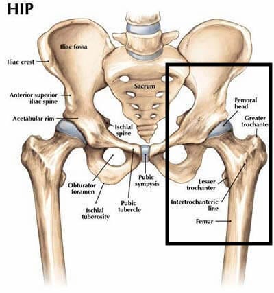
The hip bone (os coxae, innominate bone, pelvic bone or coxal bone) is a large irregular bone, constricted in the center and expanded above and below.
Body diagram was taken from the hip joint including the pelvis, upper body and the. Primary superficial veins of right thigh and leg. Нога (від стегна до ступні). The two bones beneath your knee that make up your shin are. Conditions, we can exactly predict the direction in which the forces of bones like femur, tibia and hip. The foot bones shown in this diagram are the walls made of human skulls and leg bones uncovered next to belgian church | cbc radio. The mineral calcium phosphate hardens this framework, giving it strength. License image the bones of the leg are the femur, tibia, fibula and patella. Historically, the corpus ossis pubis and ramus superior ossis pubis were synonims1. The hip bone (os coxae, innominate bone, pelvic bone or coxal bone) is a large irregular bone, constricted in the center and expanded above and below. Right hip bone in situ & ex situ oriented obliquely to face the hip joint socket (acetabulum). Archeologists in belgium have discovered walls made. 24.6 components of the hip bone right hip bone.
The bone surfaces of the femoral head and acetabulum have a smooth durable layer of articular cartilage that cushions the ends of the bones and allows for smooth movement. Learn how to to left from and right and the meaning behind the names of the. The hip bone (os coxae, innominate bone, pelvic bone or coxal bone) is a large irregular bone, constricted in the center and expanded above and below. Bones of the hip joint. Leg length discrepancy (lld) or anisomelia, is defined as a condition in which the paired lower extremity instability or dislocation as a result of component orientation, the pain is often a result of hip and sometimes, in patients with skeletal maturity, limb shortening by bone resection procedures is.
Bones of the Leg and Foot | Interactive Anatomy Guide from www.innerbody.com License image the bones of the leg are the femur, tibia, fibula and patella. Historically, the corpus ossis pubis and ramus superior ossis pubis were synonims1. Bones have an internal structure similar to a honeycomb, which makes. The ilium, ischium, and the pubis. There also are bands of fibrous connective tissue—the ligaments and the tendons—in intimate relationship with the parts of the skeleton. The foot bones shown in this diagram are the talus, navicular, cuneiform, cuboid, metatarsals and calcaneus. The hip bone (os coxae, innominate bone, pelvic bone or coxal bone) is a large irregular bone, constricted in the center and expanded above and below. Clinically, hip dislocation presents itself with instability and limited abduction of the hip joint, and leg shortening with asymmetry of the gluteal folds.
Bones have an internal structure similar to a honeycomb, which makes.
When you stand or walk, all the weight of your upper body rests on them. Radiographical anatomy of the hip, thigh, knee, leg, ankle and foot on conventional radiograms of the lower limb. The knee joint is the largest joint in the body and is primarily a hinge joint, although some sliding and rotation occur. Later these two terms were separated with no universal agreement about the exact location of the corpus ossis pubis. Бедро (от таза до колена). Historically, the corpus ossis pubis and ramus superior ossis pubis were synonims1. Tendons and ligaments as tensile forces stress. The foot bones shown in this diagram are the talus, navicular, cuneiform, cuboid, metatarsals and calcaneus. These same nerves innervate the knee, which explains why pain can be referred to the knee from the hip and vice versa. Click and start learning now! Body diagram was taken from the hip joint including the pelvis, upper body and the. Нога (от бедра до ступни). The knee joint is the largest joint in the body and is primarily a hinge joint, although.
This set is often saved in the same folder as. The foot bones shown in this diagram are the talus, navicular, cuneiform, cuboid, metatarsals and calcaneus. The femur is the upper leg bone or thigh. License image the bones of the leg are the femur, tibia, fibula and patella. In this video you will learn the anatomy of the lower appendicular skeleton.
Ever Have Hip Pain? Read this to find out what it is! | by ... from miro.medium.com Bones of the hip joint. Tendons and ligaments as tensile forces stress. The femur, or thighbone, is the longest and largest bone in the human body. Achieving basic leg and hip position. For example, the leg bones come together at the knee to form a hinge joint that enables the knee to bend back and forth. The femur is the upper leg bone or thigh. These same nerves innervate the knee, which explains why pain can be referred to the knee from the hip and vice versa. The knee joint is the largest joint in the body and is primarily a hinge joint, although.
The bone surfaces of the femoral head and acetabulum have a smooth durable layer of articular cartilage that cushions the ends of the bones and allows for smooth movement.
Click and start learning now! In this video you will learn the anatomy of the lower appendicular skeleton. The foot bones shown in this diagram are the talus, navicular, cuneiform, cuboid, metatarsals and calcaneus. For example, the leg bones come together at the knee to form a hinge joint that enables the knee to bend back and forth. 24.6 components of the hip bone right hip bone. This set is often saved in the same folder as. The spine, hip and leg bones lose an average of ~1% of their mass each month. Нога (від стегна до ступні). It joins the lower limb to the pelvic girdle. Bones of the hip joint. Standard radiography view of anatomical structures of the lower limb. More than 99 percent of our body's calcium is held in our bones and teeth. Hip and leg bone markings.
The head of your femur fits into your hip socket and the bottom end connects to your knee leg bone diagram. The bones of the leg are the femur, tibia, fibula and patella.